


Servicios Integrales
Hello everyone, today I'll share the operation procedure of the vaginal microbial immunofluorescence staining reagent.
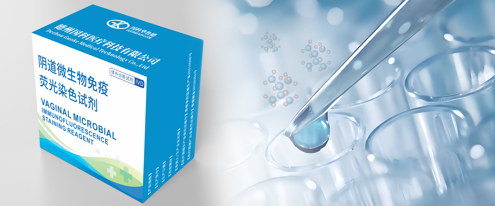
The vaginal microbial immunofluorescence kit includes reagent A and B, instruction, and a product certificate.
Reagent A is used to detect fungi in the sample.
Reagent B is used to detect pathogenic microorganisms such as epithelial cells, leukocytes, lactobacilli, bacteria, and trichomonas in the sample.
First, add one drop of vaginal microbial immunofluorescence staining reagent A to the center of area A on a glass slide.
Take a coverslip, and place one side of the coverslip in contact with the staining solution A, tilting it at a 45-degree angle and slowly flattening it to avoid air bubbles.
Use absorbent paper to remove excess solution A for better observation.
Add one drop of vaginal microbial immunofluorescence staining reagent B to the center of area B on a glass slide.
Take another coverslip, and place one side of the coverslip in contact with the staining solution B, tilting it at a 45-degree angle and slowly flattening it to avoid air bubbles.
Use absorbent paper to remove excess solution B for better observation.
The sample preparation is complete; place it on the device for observation.
If you want to learn more about microscope observation, check out our previous videos.
In the following video, we will introduce Automatic fluorescence microscopic images scanning analyzer. Thank you for watching!