


Servicios Integrales
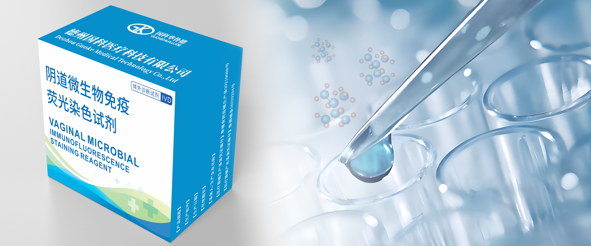
Hello everyone, today I'm going to introduce Vaginal microbial immunofluorescence staining reagent.
The vagina is an important microecological region in the female body, often home to harmless microorganisms that are beneficial and essential to health. The host body and the microbes, plus the different types of microbes themselves, all interact with each other. Some live in harmony, others keep each other in check. Together, they maintain a stable, balanced environment in the vagina. This balance is what keeps the vagina functioning normally. But if one type of microbe grows totally out of control and takes over? On the mild side, it’ll mess up the local environment. On the severe side, it can lead to vaginal inflammation.
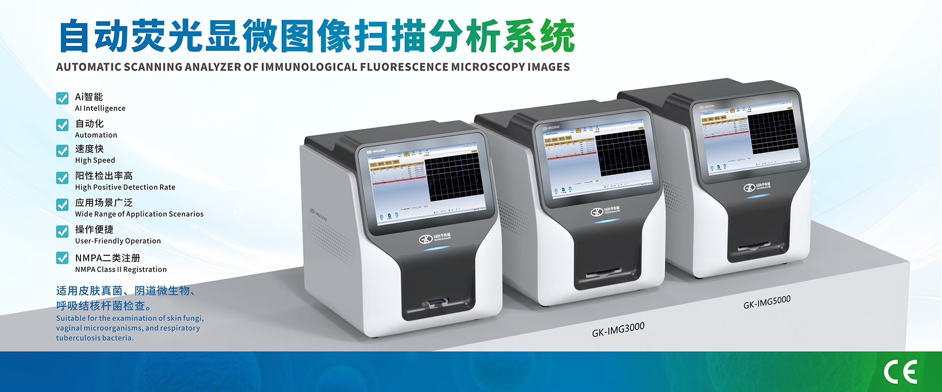
BanyanRR®Vaginal microbial immunofluorescence staining reagent can detect
pathogens related to vaginitis infections and also give doctors a clear reference for how the vaginal microecology is doing and how clean the area is.
The reagent has “fluorescently labeled antibodies”. They lock onto specific “pathogen surface antigens” in vaginal secretions and cervical exfoliated cells.Different antibody components in the reagent can respectively label β-D-glucan of fungal cell walls, lipopolysaccharide of trichomonas cell membranes, and peptidoglycan antigens of bacterial cell walls.
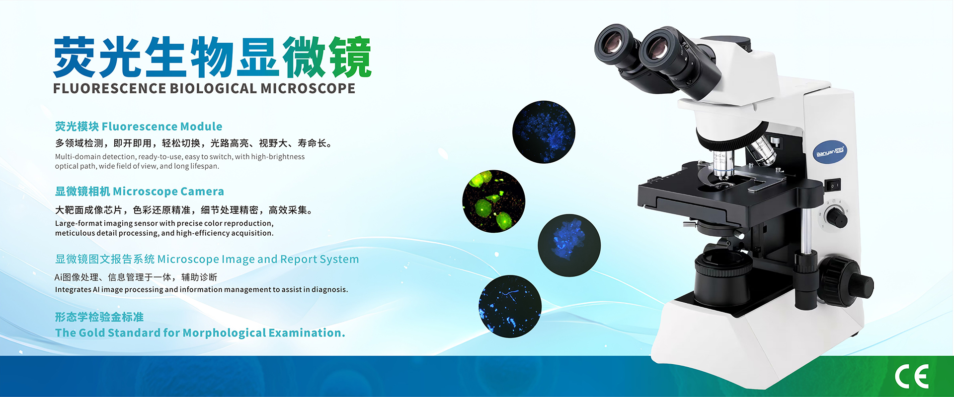
Then, we use a fluorescence microscope. When we shine two special light bands on the sample—UV band and B band —different pathogens glow with their own unique colors! Fungi light up bright blue, and trichomonas turn orange-red.
Traditional saline wet mount methods suffer from high rates of misdiagnosis and missed diagnosis. Immunofluorescence staining boosts the color brightness of target pathogens 3 times over. It also cuts the false negative rate down to under 4%.
Traditional Gram staining is not only cumbersome but also takes more than 30 minutes, while immunofluorescence staining only takes 1 minute, requires no incubation, and is simple, fast, and has significant advantages.
Immunofluorescence staining can distinguish various fungi, bacteria, and parasites, including lactobacilli, leukocytes, clue cells, Candida, and Trichomonas, among others. Target pathogens are clearly visible under fluorescent excitation.
The procedure is very simple. Even regular clinicians and junior lab techs can handle this morphological diagnosis easily.
In the next video, we'll learn about the operating procedure.Thank you for watching!